39 labelled compound microscope
(i) Draw a neat labelled ray diagram of a compound microscope. Explain ... Working: Suppose a small object AB is placed slightly away from the first focus F 0 ' of the objective lens. The objective lens forms the real, inverted and magnified image A'B', which acts as an object for eyepiece. The eyepiece is so adjusted that the image A'B' lies between the first focus F e ' and the eyepiece E. The eyepiece forms its image A'' B'' which is virtual, erect and magnified. Compound Microscope Labeled : Best Quality Product [2022] You've been seeking the right product to meet your needs, but you're unsure where to start. You require assistance in comparing items and determining which is the best - after thoroughly examining and analyzing more than 2,751 reviews products of consumers concerning the Compound microscope labeled in 2022.This blog article will help you achieve exactly that by providing reviews, buying advice ...
10 Best Compound Microscopes (Summer 2022) - The Complete Guide Best Monocular Compound Microscope: Meiji Techno MT-12 LED Monocular Compound Microscope. "If you need a monocular model, check this one that comes with a dust cover and rubber eye guards. 40x-600x magnification. Achromatic 4x, 10x, 40x, 60x lenses. LED illumination and Abbe condenser."

Labelled compound microscope
A Study of the Microscope and its Functions With a Labeled Diagram To better understand the structure and function of a microscope, we need to take a look at the labeled microscope diagrams of the compound and electron microscope. These diagrams clearly explain the functioning of the microscopes along with their respective parts. Man's curiosity has led to great inventions. The microscope is one of them. Microscope Types (with labeled diagrams) and Functions A compound microscope: Is used to view samples that are not visible to the naked eye Uses two types of lenses - Objective and ocular lenses Has a higher level of magnification - Typically up to 2000x Is used in hospitals and forensic labs by scientists, biologists and researchers to study micro organisms Compound microscope labeled diagram compound microscope parts (labeling) Flashcards | Quizlet Start studying compound microscope parts (labeling). Learn vocabulary, terms, and more with flashcards, games, and other study tools. ... connects the base to the eyepiece lens and helps to carry the microscope easily. what is 10? stage - the platform that the slides sit on. what is 11? coarse adjustment - knob that moves the slide back and ...
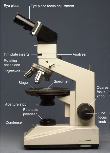
Labelled compound microscope. Draw a ray diagram of compound microscope, when final image is formed ... Draw a labelled ray diagram of an image formed by a compound microscope, when the final image lies at the least distance of distinct vision (D). ... View solution > Describe the compound microscope on the following headings: (i) Labelled ray diagram of formation of image. (ii) Magnifying power when final image is formed at least distance of ... Compound Microscope- Definition, Labeled Diagram, Principle, Parts, Uses A compound microscope is of great use in pathology labs so as to identify diseases. Various crime cases are detected and solved by drawing out human cells and examining them under the microscope in forensic laboratories. The presence or absence of minerals and the presence of metals can be identified using compound microscopes. Compound Microscope: Parts of Compound Microscope - BYJUS (A) Mechanical Parts of a Compound Microscope 1. Foot or base It is a U-shaped structure and supports the entire weight of the compound microscope. 2. Pillar It is a vertical projection. This stands by resting on the base and supports the stage. 3. Arm The entire microscope is handled by a strong and curved structure known as the arm. 4. Stage Compound Microscope – Diagram (Parts labelled), Principle and ... Feb 03, 2022 · Compound Microscope – Diagram (Parts labelled), Principle and Uses As the name suggests, a compound microscope uses a combination of lenses coupled with an artificial light source to magnify an object at various zoom levels to study the object. A compound microscope: Is used to view samples that are not visible to the naked eye
Compound Microscope - Types, Parts, Diagram, Functions and Uses Compound microscope - It has two convex lenses. It is called a compound microscope because it compounds the light as it passes through the lenses to magnify. The image of the object being viewed is enlarged because of the lens near the object. An eyepiece, an additional lens, is where real magnification takes place. Microscope Parts and Functions With Labeled Diagram and ... First, the purpose of a microscope is to magnify a small object or to magnify the fine details of a larger object in order to examine minute specimens that cannot be seen by the naked eye. Here are the important compound microscope parts... Eyepiece: The lens the viewer looks through to see the specimen. How to draw compound of Microscope easily - step by step - YouTube I will show you " How to draw compound of microscope easily - step by step "Please watch carefully and try this okay.Thanks for watching.....#microscopedrawi... Label the microscope — Science Learning Hub All microscopes share features in common. In this interactive, you can label the different parts of a microscope. Use this with the Microscope parts activity to help students identify and label the main parts of a microscope and then describe their functions. Drag and drop the text labels onto the microscope diagram.
Label Parts Of A Compound Microscope Teaching Resources | TpT This is a set of 3 tiered readings. Students will read a passage about the how to use a compound light microscope. Students will use textual evidence to answer questions and label the different parts of the microscope. It also allows students to gain prior knowledge about the compound microscope. Version A provides the most support for students. Compound Microscope Parts, Diagram Definition, Application, Working ... The compound microscopes is consists of two lenses includes, the objective lens (typically 4x, 10x, 40x or 100x) in a rotating nosepiece closer to the specimen, and the eyepiece lens (typically 10x) in the binocular eyepieces. A compound binocular microscope is more commonly used today. Labeling the Parts of the Microscope Labeling the Parts of the Microscope. This activity has been designed for use in homes and schools. Each microscope layout (both blank and the version with answers) are available as PDF downloads. You can view a more in-depth review of each part of the microscope here. Microscope Parts, Function, & Labeled Diagram - slidingmotion Microscope parts labeled diagram gives us all the information about its parts and their position in the microscope. Microscope Parts Labeled Diagram The principle of the Microscope gives you an exact reason to use it. It works on the 3 principles. Magnification Resolving Power Numerical Aperture. Parts of Microscope Head Base Arm Eyepiece Lens
Mikroskop Nedir? Nasıl Kullanılır? | FenEhli.com
Compound Microscope: Definition, Diagram, Parts, Uses ... The compound microscope is mainly used for studying the structural details of cell, tissue, or sections of organs. The parts of a compound microscope can be classified into two: Non-optical parts Optical parts Non-optical parts Base The base is also known as the foot which is either U or horseshoe-shaped.

Simple Columnar Epithelium: Description: Single layer of elongated ...
Parts of a Compound Microscope and Their Functions Compound microscope magnification is determined by multiplying the eyepiece and objective powers. When viewed through a 5X eyepiece with a 10X objective, an item is magnified 5 x 10=50 times. The magnification is 10 x 45 = 450 times when using a 10X eyepiece and a 45X objective. How to Use the Compound Microscope

Labeling the parts of the microscope. (Blank diagram available for ...
Parts of a microscope with functions and labeled diagram There are three structural parts of the microscope i.e. head, base, and arm. Head - This is also known as the body. It carries the optical parts in the upper part of the microscope. Base - It acts as microscopes support. It also carries microscopic illuminators.
Optical Microscopy and Specimen - Using the Transmission Microscope
Compound Microscope Parts – Labeled Diagram and their ... The term "compound" refers to the microscope having more than one lens. Basically, compound microscopes generate magnified images through an aligned pair of the objective lens and the ocular lens. In contrast, "simple microscopes" have only one convex lens and function more like glass magnifiers.

Compound Light Microscope
(a) Draw a labelled ray diagram of compound microscope, when final ... (a) Draw a labelled ray diagram of compound microscope, when final image forms at the least distance of distinct vision. (b) Why is its objective of short focal length and of short aperture, compared to its eyepiece? Explain. (c) The focal length of the objective is 4 cm while that of eyepiece is 10 cm. The object is placed at a distance of 6 cm from the objective lens.
Quiz: Microscope Basics by Cynthia Zack | Teachers Pay Teachers
Compound Microscope Labeled Diagram | Quizlet QUESTION. The total magnification of a specimen being viewed with a 10X ocular lens and a 40X objective lens is. 15 answers. QUESTION. a mosquito beats its wings up and down 600 times per second, which you hear as a very annoying 600 Hz sound. if the air outside is 20 C, how far would a sound wave travel between wing beats. 2 answers.

This is an example of a Eukaryotic cell under a microscope
Compound Microscope Parts, Functions, and Labeled Diagram Nov 18, 2020 · Compound Microscope Definitions for Labels. Eyepiece (ocular lens) with or without Pointer: The part that is looked through at the top of the compound microscope. Eyepieces typically have a magnification between 5x & 30x. Monocular or Binocular Head: Structural support that holds & connects the eyepieces to the objective lenses.

Deaf Scientist Corner
(b) Why both objective and eyepiece of a compound microscope must have ... (a) Draw the labelled ray diagram for the formation of image by a compound microscope. Derive an expression for its total magnification (or magnifying power), when the final image is formed at the near point. (b) Why both objective and eyepiece of a compound microscope must have short focal lengths?
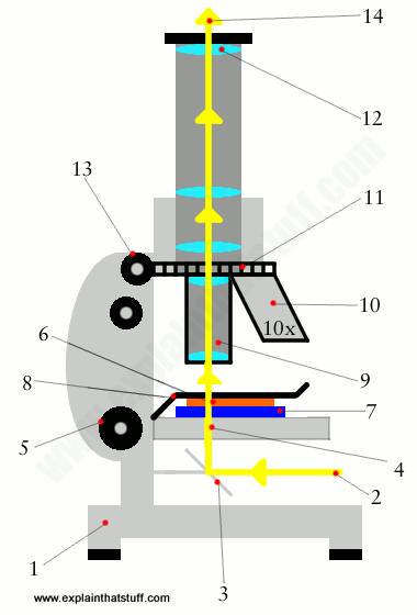
How does a microscope work? - Explain that Stuff
(a) Draw a labelled ray diagram of a compound microscope. (b) Derive an ... selected May 26, 2018 by Vikash Kumar Best answer (a) Labelled diagram of compound microscope. The objective lens form image A' B' near the first focal point ofeyepiece. (b) Angular magnification of objective lens m0 = linear magnification h'/h where L is the distance between second focal point of the objective and first focal point of eyepiece.
Post a Comment for "39 labelled compound microscope"