41 microscope diagram labeled
PDF Parts of the Light Microscope - Science Spot B. NOSEPIECE microscope when carried Holds the HIGH- and LOW- power objective LENSES; can be rotated to change MAGNIFICATION. Power = 10 x 4 = 40 Power = 10 x 10 = 100 Power = 10 x 40 = 400 What happens as the power of magnification increases? Labelled Diagram of Compound Microscope - Biology Discussion The below mentioned article provides a labelled diagram of compound microscope. Part # 1. The Stand: The stand is made up of a heavy foot which carries a curved inclinable limb or arm bearing the body tube. The foot is generally horse shoe-shaped structure (Fig. 2) which rests on table top or any other surface on which the microscope in kept.
Compound Microscope Labeled Diagram | Quizlet QUESTION. The total magnification of a specimen being viewed with a 10X ocular lens and a 40X objective lens is. 15 answers. QUESTION. a mosquito beats its wings up and down 600 times per second, which you hear as a very annoying 600 Hz sound. if the air outside is 20 C, how far would a sound wave travel between wing beats. 2 answers.

Microscope diagram labeled
Parts of Stereo Microscope (Dissecting microscope) - labeled diagram ... Labeled part diagram of a stereo microscope Major structural parts of a stereo microscope There are three major structural parts of a stereo microscope. The viewing Head includes the upper part of the microscope, which houses the most critical optical components, including the eyepiece, objective lens, and light source of the microscope. Parts of a microscope with functions and labeled diagram Figure: Diagram of parts of a microscope There are three structural parts of the microscope i.e. head, base, and arm. Head - This is also known as the body. It carries the optical parts in the upper part of the microscope. Base - It acts as microscopes support. It also carries microscopic illuminators. Microscope Labeling - The Biology Corner Microscope Labeling. This simple worksheet pairs with a lesson on the light microscope, where beginning biology students learn the parts of the light microscope and the steps needed to focus a slide under high power. The labeling worksheet could be used as a quiz or as part of direct instruction where students label the microscope as you go ...
Microscope diagram labeled. Compound Microscope- Definition, Labeled Diagram, Principle, Parts, Uses The naked eye can now view the specimen at magnification 400 times greater and so microscopic details are revealed. Alternatively, the magnification of the compound microscope is given by: m = D/ fo * L/fe where, D = Least distance of distinct vision (25 cm) L = Length of the microscope tube fo = Focal length of the objective lens Compound Microscope Parts, Functions, and Labeled Diagram Compound Microscope Parts, Functions, and Labeled Diagram Parts of a Compound Microscope Each part of the compound microscope serves its own unique function, with each being important to the function of the scope as a whole. Microscope Diagram Labeled, Unlabeled and Blank | Parts of a Microscope ... This PDF contains the following: 1. Parts of a Microscope Diagram - Color 2. Parts of a Microscope Diagram - Black and White 3. Blank Parts of a Microscope Diagram - Black and White 4. Blank, Unlabeled Parts of a Microscope Diagram - Black and White 5. Blank Parts of a Microscope Diagram - Color 6. Blank, Unlabeled… Binocular Microscope Anatomy - Parts and Functions with a Labeled Diagram Now, I will discuss the details anatomy of the light compound microscope with the labeled diagram. Why it is called binocular: because it has two ocular lenses or an eyepiece on the head that attaches to the objective lens, this ocular lens magnifies the image produced by the objective lens. Binocular microscope parts and functions
Microscope Parts and Functions With Labeled Diagram and Functions How ... Most specimens are mounted on slides, flat rectangles of thin glass. The specimen is placed on the glass and a cover slip is placed over the specimen. This allows the slide to be easily inserted or removed from the microscope. It also allows the specimen to be labeled, transported, and stored without damage. Parts of the Microscope with Labeling (also Free Printouts) 5. Knobs (fine and coarse) By adjusting the knob, you can adjust the focus of the microscope. The majority of the microscope models today have the knobs mounted on the same part of the device. Image 5: The circled parts of the microscope are the fine and coarse adjustment knobs. Picture Source: bp.blogspot.com. Microscope Labeling Practice Diagram | Quizlet Where the microscope slide is placed for viewing. Coarse Focus Knob (Coarse Adjustment) Elevates or lowers the stage a large distance with each turn of the knob. ... Anatomy and Physiology 46 Body Region Terms. 46 terms. AngieTepile. Sets with similar terms. Parts of a Microscope. 15 terms. joshferguson1. Microscope Parts and Functions. Hair Under a Microscope - AnatomyLearner All the following microscopic features are well identified in the hair-labeled diagram. Epidermis and dermis layer of the skin with hair, Different parts of a hair shaft - medulla, cortex, and cuticle, Hair root in the epidermis or hypodermis of the animal's skin, Connective tissue layer that surrounds the hair shaft or follicles,
Microscope Diagram Labeled, Unlabeled and Blank | Parts of a Microscope ... This PDF contains the following: 1. Parts of a Microscope Diagram - Color 2. Parts of a Microscope Diagram - Black and White 3. Blank Parts of a Microscope Diagram - Black and White 4. Blank, Unlabeled Parts of a Microscope Diagram - Black and White 5. Blank Parts of a Microscope Diagram - Color 6. Blank, Unlabeled… Microscope, Microscope Parts, Labeled Diagram, and Functions Microscope, Microscope Parts, Labeled Diagram, and Functions What is Microscope? A microscope is a laboratory instrument used to examine objects that are too small to be seen by the naked eye. It is derived from Ancient Greek words and composed of mikrós, "small" and skopeîn,"to look" or "see". Label Microscope Diagram - EnchantedLearning.com diaphragm - an adjustable opening under the stage, allowing different amounts of light onto the stage. eyepiece - where you place your eye. fine focus adjustment - a knob that makes small adjustments to the focus (it is often smaller than the coarse focus knob). high-power objective - a large lens with high magnifying power. Microscope Parts, Function, & Labeled Diagram - slidingmotion Microscope parts labeled diagram gives us all the information about its parts and their position in the microscope. Microscope Parts Labeled Diagram The principle of the Microscope gives you an exact reason to use it. It works on the 3 principles. Magnification Resolving Power Numerical Aperture. Parts of Microscope Head Base Arm Eyepiece Lens
Skin (Integumentary System)
PDF Parts of a Microscope Printables - Homeschool Creations Label the parts of the microscope. You can use the word bank below to fill in the blanks or cut and paste the words at the bottom. Microscope Created by Jolanthe @ HomeschoolCreations.net. Parts of a eyepiece arm stageclips nosepiece focusing knobs illuminator stage objective lenses

cell and organelles Dr.Jastrow's electron microscopic atlas
Compound Microscope Parts - Labeled Diagram and their Functions - Rs ... Labeled diagram of a compound microscope Major structural parts of a compound microscope There are three major structural parts of a compound microscope. The head includes the upper part of the microscope, which houses the most critical optical components, and the eyepiece tube of the microscope.
Object Segmentation using Fuzzy Divergence in python: A case study over ...
Microscope Labeling - The Biology Corner Microscope Labeling. This simple worksheet pairs with a lesson on the light microscope, where beginning biology students learn the parts of the light microscope and the steps needed to focus a slide under high power. The labeling worksheet could be used as a quiz or as part of direct instruction where students label the microscope as you go ...
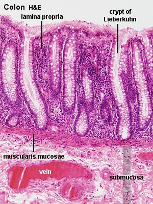
Gastrointestinal Tract - Colon Histology - Embryology
Parts of a microscope with functions and labeled diagram Figure: Diagram of parts of a microscope There are three structural parts of the microscope i.e. head, base, and arm. Head - This is also known as the body. It carries the optical parts in the upper part of the microscope. Base - It acts as microscopes support. It also carries microscopic illuminators.
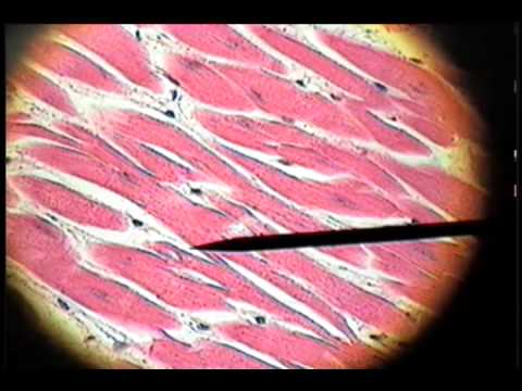
Slide 35 - Cardiac Muscle - YouTube
Parts of Stereo Microscope (Dissecting microscope) - labeled diagram ... Labeled part diagram of a stereo microscope Major structural parts of a stereo microscope There are three major structural parts of a stereo microscope. The viewing Head includes the upper part of the microscope, which houses the most critical optical components, including the eyepiece, objective lens, and light source of the microscope.

bread mold (Rhizopus stolonifer ) on cultivated garlic (Allium sativum ...
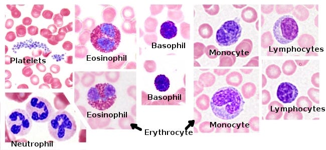
Tissues Flashcards | Easy Notecards

Quia - Protist Vocabulary

small intestine histology labeled | Medical school motivation, Human ...
Post a Comment for "41 microscope diagram labeled"