42 heart diagram without labels
Circulatory System Diagram - New Health Advisor This circuit typically includes the movement of blood inside heart and 'myocardium' (the membrane of heart). Coronary circuit mainly consists of cardiac veins including anterior cardiac vein, small vein, middle vein and great (large) cardiac vein. There are different types of circulatory system diagrams; some have labels while others don't. Human Heart Diagram Labeled - Science Trends List Of Heart Structures Heart Chambers Ventricles - The bottom two heart chambers. Atra - The upper two heart chambers. Wall Of The Heart Sinoatrial Node - A collection of tissue that releases electrical impulses and defines the rate of contraction for the heart. Atrioventricular Bundle - The fibers which transmit cardiac impulses.
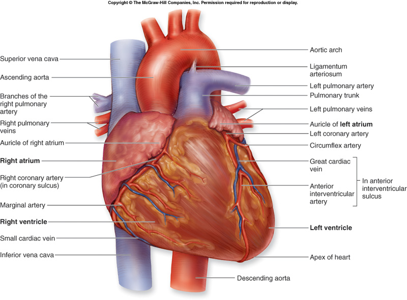
A Labeled Diagram of the Human Heart You Really Need to See The human heart, comprises four chambers: right atrium, left atrium, right ventricle and left ventricle. The two upper chambers are called the left and the right atria, and the two lower chambers are known as the left and the right ventricles. The two atria and ventricles are separated from each other by a muscle wall called 'septum'.

Heart diagram without labels
Heart Anatomy: Labeled Diagram, Structures, Blood Flow ... - EZmed There are 4 chambers, labeled 1-4 on the diagram below. To help simplify things, we can convert the heart into a square. We will then divide that square into 4 different boxes which will represent the 4 chambers of the heart. The boxes are numbered to correlate with the labeled chambers on the cartoon diagram. Blank Heart Diagrams Diagram Link - Heart Diagram Drawing | Human heart ... All the best Heart Diagram Drawing 36+ collected on this page. Feel free to explore, study and enjoy paintings with PaintingValley.com C Caroline Kong 98 followers More information Blank Heart Diagrams Diagram Link - Heart Diagram Drawing Find this Pin and more on Study Buddy by Caroline Kong. Simple Heart Diagram Human Heart Diagram 13+ Heart Diagram Templates - Sample, Example, Format Download Free heart diagrams can be helpful for students in understanding the heart and its functioning. Human heart is a complicated figure and for students from science, they will often need the images of the heart for its illustration. The above collection of heart samples will make it easier for students to download, print and use it in their projects.
Heart diagram without labels. Label Heart Anatomy Diagram Printout - EnchantedLearning.com Every day, the heart pumps about 2,000 gallons (7,600 liters) of blood, beating about 100,000 times. Label the heart anatomy diagram below using the heart glossary. Note: On the diagram, the right side of the heart appears on the left side of the picture (and vice versa) because you are looking at the heart from the front. Enchanted Learning Search Human Heart Diagram Without Labels - Labelling Worksheet It's also made up of four valves - these are known as the tricuspid, pulmonary, mitral and aortic valves. With this heart diagram without labels, you can familiarise your students with all the correct terms and help them recognise all these features of the anatomy. The above video may be from a third-party source. Heart Blood Flow | Simple Anatomy Diagram, Cardiac Circulation ... - EZmed Step 1 involves blood vessels, similar to what we saw with step 1 in the right side of the heart. The pulmonary veins carry oxygenated blood from the lungs to the left side of the heart, specifically the left atrium. There will be better images of the pulmonary veins shown in the images later in this post. 2. Left Atrium The Anatomy of the Heart - Quiz 1 - Free Anatomy Quiz The heart - an image of the heart with blank labels attached The circulatory system - upper body image, with blank labels attached The circulatory system - lower body image, with blank labels attached The circulatory system - a PDF file of the upper and lower body for printing out to use off-line Articles :
Ear Diagram Unlabeled - Wiring Diagrams Ear Diagram Unlabeled. Worksheet asks children to label the parts of the ear and re-arrange a jumbled up Carnivores, omnivores and herbivores Venn diagram. Students knowledge of unlabelled diagram of the human ear diagrams; diagram heart diagrams. Knowledge of biology human lung erica l. Heart Labeling Quiz: How Much You Know About Heart Labeling? Here is a Heart labeling quiz for you. The human heart is a vital organ for every human. The more healthy your heart is, the longer the chances you have of surviving, so you better take care of it. Take the following quiz to know how much you know about your heart. Questions and Answers 1. What is #1? 2. What is #2? 3. What is #3? 4. What is #4? Human Heart Diagram Without Labels - Pinterest ABOUT THIS ACTIVITY: Illustrates the pathway of blood through the heart. The areas of the heart with MORE oxygen are labeled with an "R". Students will color these areas RED. The areas of the heart with LESS oxygen are labeled with a "B". Students will color these areas BLUE. Heart Diagram - 15+ Free Printable Word, Excel, EPS, PSD Template ... Heart Diagram - 15+ Free Printable Word, Excel, EPS, PSD Template Download A heart diagram is a popular design used by different people for various uses. It can be used by a teacher or student for academic purpose, by a friend or relative for mutually sending and exchanging cards or for baby toys or printing on dresses etc.
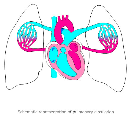
Label the heart — Science Learning Hub In this interactive, you can label parts of the human heart. Drag and drop the text labels onto the boxes next to the heart diagram. If you want to redo an answer, click on the box and the answer will go back to the top so you can move it to another box. If you want to check your answers, use the Reset Incorrect button. Heart Diagram Unlabeled - Cliparts.co Heart Diagram Unlabeled 84 images of Heart Diagram Unlabeled. You can use these free cliparts for your documents, web sites, art projects or presentations. Don't forget to link to this page for attribution! Diagram of Blood Flow Through the Heart - Bodytomy The deoxygenated blood from the heart enters the lungs through the pulmonary valve as seen in the human heart diagram. This process is called pulmonary circulation. From the pulmonic valve, the blood travels to the pulmonary artery into the tiny capillary vessels of the lungs. The oxygen present in the tiny air sacs enters the blood through the ... Cardiovascular system: Diagrams, quizzes, free worksheets - Kenhub Cardiovascular system diagrams, quizzes and free worksheets. The cardiovascular system is a vital organ system which is quite literally at the centre of everything. Comprised of the heart, blood vessels and the blood itself, it is divided into two loops which both begin in the heart. The pulmonary circuit is responsible for exchanging blood ...

13 Best Images of Hip Anatomy Of The Worksheet - Sunflower Anatomy ...
The Human Heart Cardiovascular System Labeling Worksheet This handy heart worksheet gives your children the opportunity to show how much they've learned about this topic. Using the blank heart diagram students are asked to label the aorta, superior vena cava, pulmonary arteries, pulmonary veins, atrium, ventricles, and aortic valves. This simple human heart diagram could be used as both a starter or plenary in order to assess students ...
Free Blank Heart Diagram, Download Free Blank Heart Diagram png images ...
Draw The Venn Diagram Of A-B : Heart Diagram With Labels â€" Heart ... Heart Diagram With Labels â€" Heart Diagram Without Labels from 5 draw appropriate venn diagram for each of the following : Just like with numbers, we use parentheses if . We have to draw diagram for complement of (a ⋃ b) i.e.(a ∪ b) . The following pages on the english wikipedia use this file (pages .
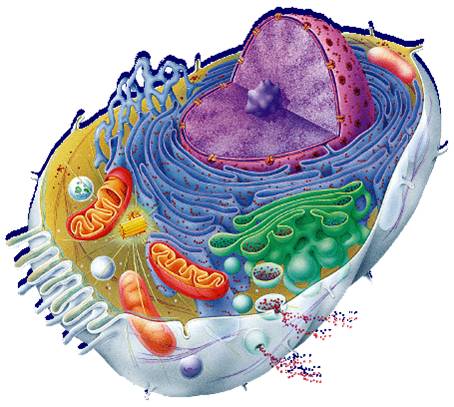
Free Animal Cell Unlabeled, Download Free Clip Art, Free Clip Art on ...
Labelling the heart — Science Learning Hub Blood transports oxygen and nutrients to the body. It is also involved in the removal of metabolic wastes. In this interactive, you can label parts of the human heart. Drag and drop the text labels onto the boxes next to the diagram. Selecting or hovering over a box will highlight each area in the diagram.
Circulatory System Diagram - Cardiovascular System and Blood ...
Diagram of Human Heart and Blood Circulation in It The outermost layer of your heart wall is called the epicardium, which is basically a very thin layer of serous membrane. The membrane provides lubrication and protection to the outer side of your heart, as you can see in heart diagram labeled. Myocardium. Right beneath epicardium is another relatively thicker layer called myocardium.
What purpose do heart valves serve? - Quora
Human Heart (Anatomy): Diagram, Function, Chambers, Location in Body Chambers of the Heart. The heart is a muscular organ about the size of a fist, located just behind and slightly left of the breastbone. The heart pumps blood through the network of arteries and ...
The Anatomy and Physiology of Animals/Circulatory System Worksheet ...
circulatory system worksheet without labels - Google Search | Heart ... Heart Diagram Nursing School Notes The normal Adult Human Heart Weighs 250 to 350g. It has 4 chambers (Two Atria and Two Ventricles-both left and right). How does it function? Why is it so important to the Human physiology? A Andrew Rudin MD Heart Health | Andrew Rudin MD

Free Unlabeled Eye Diagram, Download Free Unlabeled Eye Diagram png ...
Heart Diagram with Labels and Detailed Explanation - BYJUS Well-Labelled Diagram of Heart. The heart is made up of four chambers: The upper two chambers of the heart are called auricles. The lower two chambers of the heart are called ventricles. The heart wall is made up of three layers: The outer layer of the heart wall is called epicardium. The middle layer of the heart wall is called myocardium.
Circulatory System Diagram - Cardiovascular System and Blood ...
How to Draw a Human Heart: 11 Steps (with Pictures) - wikiHow 3. Sketch a forked tube extending from the top of the rounded bump. To make the superior vena cava, draw a tube coming from the top of the right atrium. Make the tube fork about the same length as the bump you made for the right atrium chamber. Blood enters the right atrium through the superior vena cava.
Post a Comment for "42 heart diagram without labels"