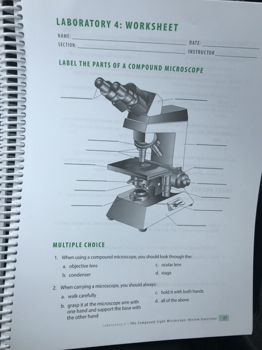
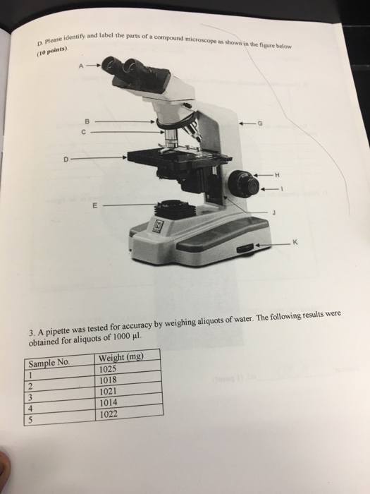
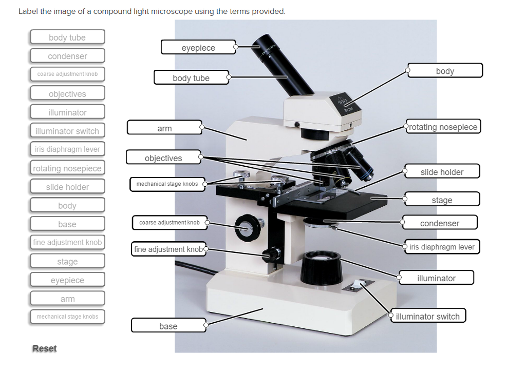
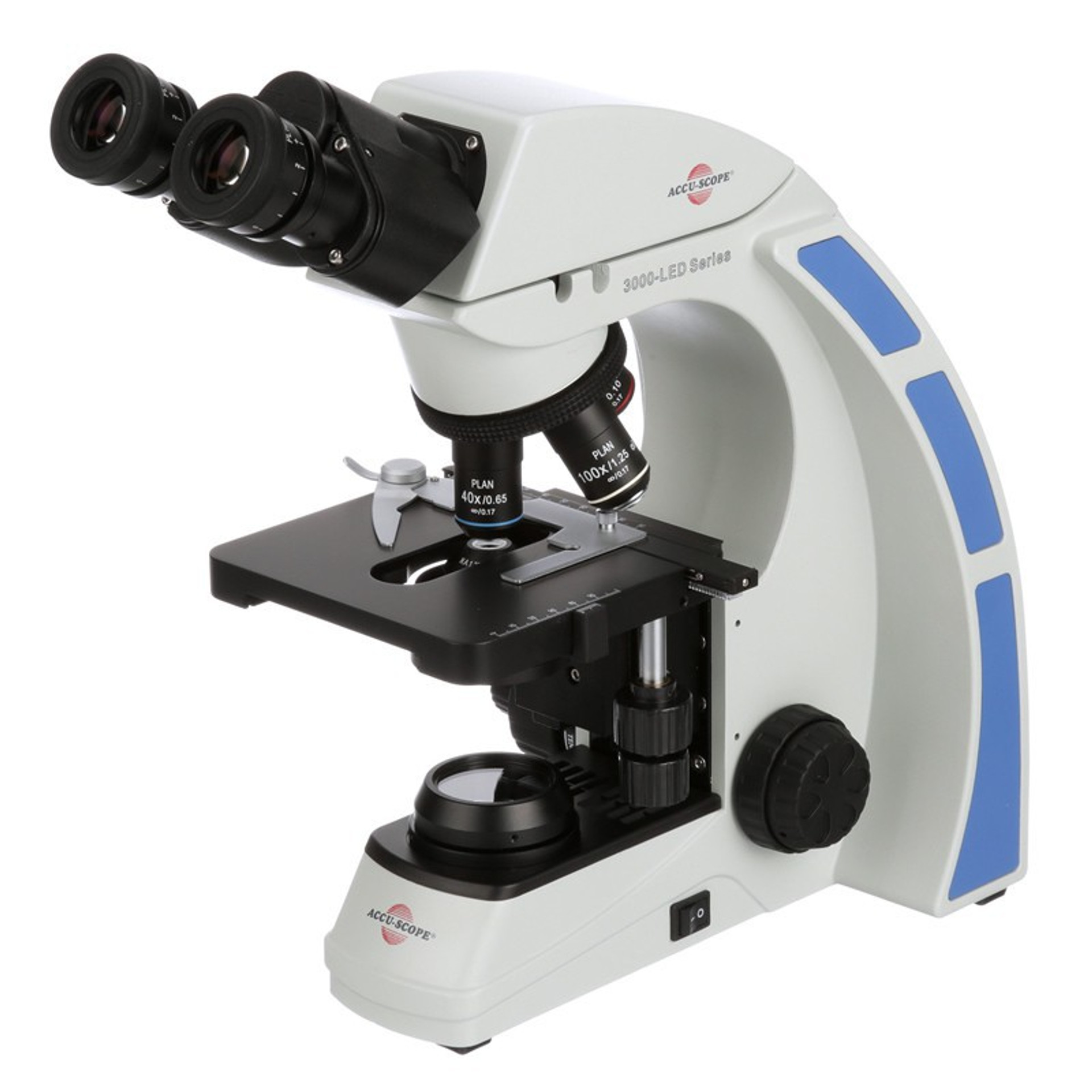
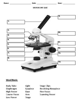
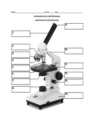
40 label the parts of the compound microscope
Microscope Diagram - slide preparation biology4isc, anthrax microbewiki ... Microscope Diagram - 15 images - lycopodium, cell division of e coli with continuous media flow youtube, labelled microscope diagram gcse micropedia, give a well labelled diagram of compound microscope using of typical, How To Prepare A Light Microscope | Americanwarmoms.org Molecular Expressions Science Optics And You Intel Play Qx3 Computer Microscope Samples For Polarized Light Microscopy
www1.udel.edu › biology › ketchamUD Virtual Compound Microscope - University of Delaware ©University of Delaware. This work is licensed under a Creative Commons Attribution-NonCommercial-NoDerivs 2.5 License.Creative Commons Attribution-NonCommercial-NoDerivs 2

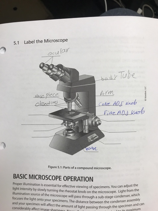
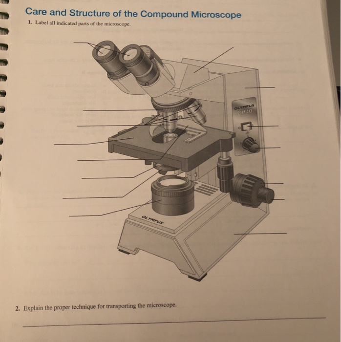
Label the parts of the compound microscope
researchtweet.com › microscope-parts-labeledMicroscope, Microscope Parts, Labeled Diagram, and Functions Jan 19, 2022 · Illuminator: Illuminator is the most important microscope parts and it serve as light source for a microscope during slide specimen visualization. It is a continuous source of light (110 volts) used in place of a mirror. The mirror of microscope is used to reflect light from the external light source up through the bottom of the stage. Microorganisms | Free Full-Text | Efficacy of Off-Label Anti-Amoebic ... Of the eight off-label anti-amoebic drugs tested, only PVPI allowed for a complete suppression of trophozoite formation by drug-challenged cysts for all four Acanthamoeba isolates in all five biological replicates. ... Bright-field images were taken with a Leica DMI4000 B microscope every week for up to 3 weeks with a 10-fold objective and 8 ... Motility Test (Theory) - Amrita Vishwa Vidyapeetham Virtual Lab Fig:-Lophotrichous flagellum seen under light microscope . Flagella are spread fairly evenly over the whole surface of peritrichous bacteria . Fig:-Peritrichous flagellum seen under light microscope . When anticlockwise rotation is resumed, the cell tends to move in a new direction. This ability is important, since it allows bacteria to change ...
Label the parts of the compound microscope. Microscope Answers Worksheet Pre Lab Part 1; Part 2; Part 3; Part 4; Part 5; Part 6; ... any type of subject material you will not come across at any place else for you to become familiar with the use of the compound light microscope to better understand cell biology works both ways answers e2020 answers for evolution lab 37 answer key exploring science 7' 'Love S Masquerade ... Under Microscope Elodea The Search: Elodea Under The Microscope. · During the observations under 40X magnification, the horizontal and lateral sizes of the Elodea cells were calculated, according to the calculations in epithelial cell samples under 40X Exercise 1: Plant Cells Making a Temporary Wet Mount of Elodea (water plant) 1 Record your observations Place a small, thin piece of the elodea, onion and cork on one ... Quizlet Parts And Use Worksheet Microscope And the parts to make it cost less than a dollar Base: The base of a compound microscope is helps in supporting the microscope and contains the illuminator When you are finished using the microscope: a when transporting the microscope, hold it in an upright position with one hand on its arm and the other supporting its base Literal or literally ... sciencing.com › difference-between-compoundDifference Between Compound & Dissecting Microscopes Mar 10, 2018 · Dissecting and compound light microscopes are both optical microscopes that use visible light to create an image. Both types of microscope magnify an object by focusing light through prisms and lenses, directing it toward a specimen, but differences between these microscopes are significant.
ECLIPSE Ti2 Series | Inverted Microscopes - Nikon Instruments Inc. The ECLIPSE Ti2 inverted microscope delivers an unparalleled 25mm field of view (FOV) that revolutionizes the way you see. With this incredible FOV, the Ti2 maximizes the sensor area of large-format CMOS cameras without making compromises, and significantly improves data throughput. The Ti2's exceptionally stable, drift-free platform is ... Lichen - Wikipedia A lichen (/ ˈ l aɪ k ən / LY-kən, also UK: / ˈ l ɪ tʃ ən / LITCH-ən) is a composite organism that arises from algae or cyanobacteria living among filaments of multiple fungi species in a mutualistic relationship. Lichens have properties different from those of their component organisms. They come in many colors, sizes, and forms and are sometimes plant-like, but are not plants. They ... www1.udel.edu › biology › ketchamMicroscopy Pre-lab Activities - University of Delaware Microscope controls: turn knobs (click and hold on upper or lower portion of knob) throw switches (click and drag) turn dials (click and drag) move levers (click and drag) changes lenses (click and drag on objective housing) select a specimen (click on a slide) scheme work biology - Free KCPE Past Papers Organisms in the school compound; ... Draw and label the light microscope; Description of a cell; Drawing and labeling the light microscope . ... Golden tips Biology Page 15-16; KLB teachers book 1 pages 23-25 . 10. 1-2. THE CELL. Parts of the light microscope and their functions . Calculation of magnification using light microscope. By the end ...
› p-3470-what-is-aWhat is a Compound Microscope? | Microscope World Blog A compound microscope is a high power (high magnification) microscope that uses a compound lens system. A compound microscope has multiple lenses: the objective lens (typically 4x, 10x, 40x or 100x) is compounded (multiplied) by the eyepiece lens (typically 10x) to obtain a high magnification of 40x, 100x, 400x and 1000x. C3s2 Compound Name Chegg - vej.mondo.vi.it Search: C3s2 Compound Name Chegg. See if the charges are balanced (if they are you're done!) Add subscripts (if necessary) so the charge for the entire compound is zero So if the formula has hydrogen written first, then this usually indicates that the hydrogen is an H+ cation and that the compound is an acid Naming Compounds 2 While there are millions of organic compounds, there is a fairly ... Metaphase - Genome.gov Metaphase is a stage during the process of cell division (mitosis or meiosis). Normally, individual chromosomes are spread out in the cell nucleus. During metaphase, the nucleus dissolves and the cell's chromosomes condense and move together, aligning in the center of the dividing cell. At this stage, the chromosomes are distinguishable when ... Tissue distribution and metabolic profiling of cyclosporine (CsA) in ... Therapeutic peptides are a fast-growing class of pharmaceuticals. Like small molecules, the costs associated with their discovery and development are significant. In addition, since the preclinical data guides first-in-human studies, there is a need for analytical techniques that accelerate and improve our understanding of the absorption, distribution, metabolism, and excretion (ADME ...
Basic Microscope Diagram - microscope diagram purposegames, images 01 ... Basic Microscope Diagram - 15 images - label the neuron clip art at vector clip art online, microscope diagram fill online printable fillable blank pdffiller, animal anatomy biology4isc, images 01 introduction and terminology basic human anatomy,
Mr. Jones's Science Class Matter: Atoms and Properties - Open Response Question 3. Force and Motion - Open Response Question 3. Forms of Energy - Open Response Question 1. Forms of Energy - Open Response Question 2. Earth's Structure & Natural Processes - Open Response Question 1.
Paramecium Classification Structure Function And Characteristics Cell Nucleus - function, structure, and under a microscope. The nucleus is a specialized organelle that contains double-layer membranes with pores. The main function of the nucleus is to govern cell activities and to carry genetic information to pass to the next generation.
Cell Structure (Learn) : Biology : Class 9 : Amrita Vidyalayam ... This was in the year 1665 when Hooke made this chance observation through a self-designed microscope. Robert Hooke called these boxes cells. Cell is a Latin word for 'a little room'. This may seem to be a very small and insignificant incident but it is very important in the history of science. This was the very first time that someone had ...
› anton-van-leeuwenhoek-1991633Antonie van Leeuwenhoek, Father of Microbiology - ThoughtCo Jul 21, 2019 · Also credited with the invention of the microscope about the same time was Hans Lippershey, the inventor of the telescope. Their work led to others' research and development on telescopes and the modern compound microscope, such as Galileo Galilei, Italian astronomer, physicist, and engineer whose invention was the first given the name ...
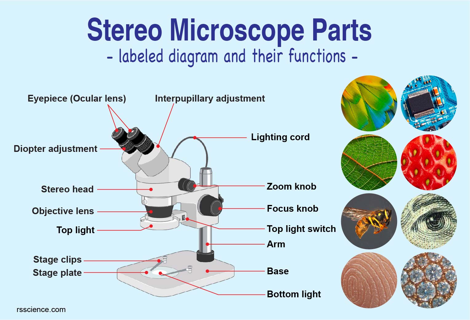
microbenotes.com › parts-of-a-microscopeParts of a microscope with functions and labeled diagram Apr 19, 2022 · Figure: Diagram of parts of a microscope. There are three structural parts of the microscope i.e. head, base, and arm. Head – This is also known as the body. It carries the optical parts in the upper part of the microscope. Base – It acts as microscopes support. It also carries microscopic illuminators.
Differences between Light Microscope and Electron Microscope Light Microscope. Electron Microscope. Illuminating source is the Light. Illuminating source is the beam of electrons. Specimen preparation takes usually few minutes to hours. Specimen preparation takes usually takes few days. Live or Dead specimen may be seen. Only Dead or Dried specimens are seen. Condenser, Objective and eye piece lenses are ...
What Is The Purpose Of A Microscope - facit.edu.br Sep 29, 2020 . Fundus photography is the process of taking serial photographs of the interior of your eye through the pupil. A fundus camera is a specialized low-power microscope attached to a camera used to examine structures such as the optic disc, retina, and lens..
Light Microscope (Theory) - Amrita Vishwa Vidyapeetham The modern compound microscope consists of two lens system, the objective and the ocular or eye piece. The first magnified image obtained with objective lens, is again magnified by the eye piece to give a virtual inverted image. The total magnification the product of the magnifications of two lens systems. Parts of a Microscope . It consists of ...
Motility Test (Theory) - Amrita Vishwa Vidyapeetham Virtual Lab Fig:-Lophotrichous flagellum seen under light microscope . Flagella are spread fairly evenly over the whole surface of peritrichous bacteria . Fig:-Peritrichous flagellum seen under light microscope . When anticlockwise rotation is resumed, the cell tends to move in a new direction. This ability is important, since it allows bacteria to change ...
Microorganisms | Free Full-Text | Efficacy of Off-Label Anti-Amoebic ... Of the eight off-label anti-amoebic drugs tested, only PVPI allowed for a complete suppression of trophozoite formation by drug-challenged cysts for all four Acanthamoeba isolates in all five biological replicates. ... Bright-field images were taken with a Leica DMI4000 B microscope every week for up to 3 weeks with a 10-fold objective and 8 ...
researchtweet.com › microscope-parts-labeledMicroscope, Microscope Parts, Labeled Diagram, and Functions Jan 19, 2022 · Illuminator: Illuminator is the most important microscope parts and it serve as light source for a microscope during slide specimen visualization. It is a continuous source of light (110 volts) used in place of a mirror. The mirror of microscope is used to reflect light from the external light source up through the bottom of the stage.










Post a Comment for "40 label the parts of the compound microscope"