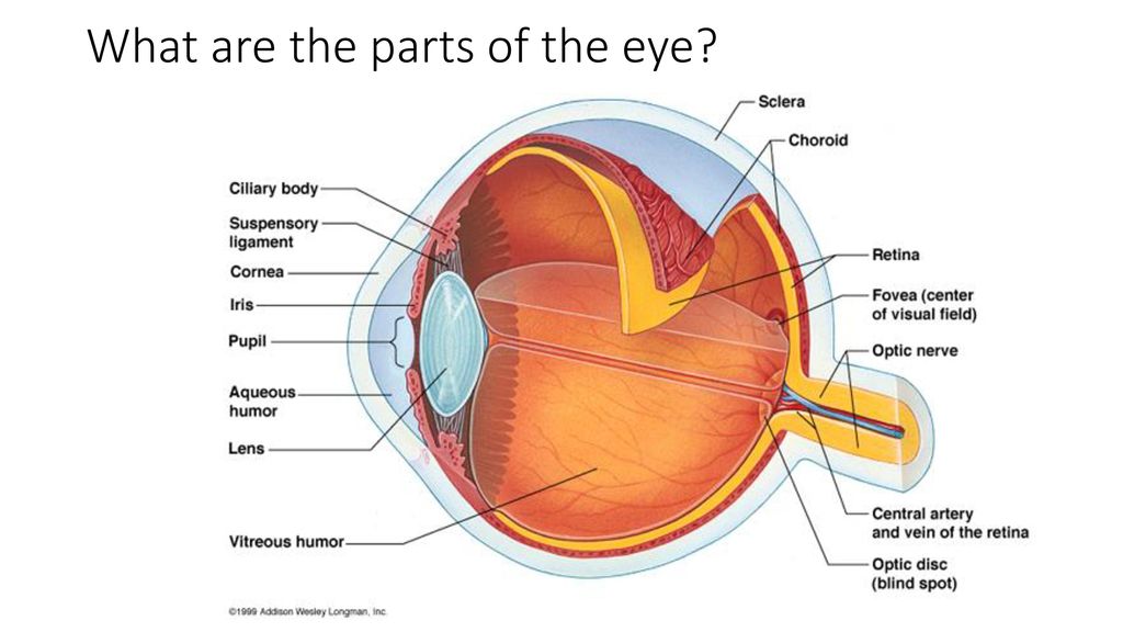
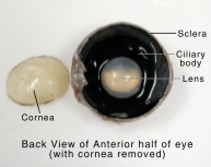
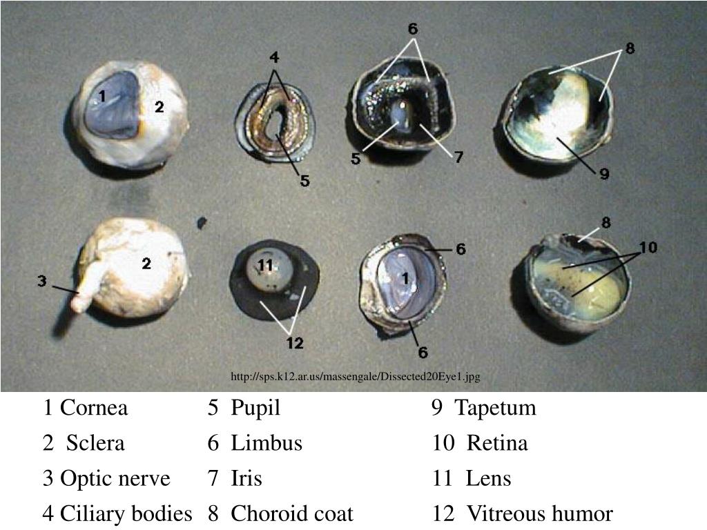
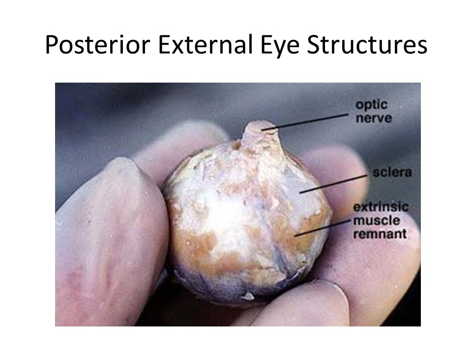
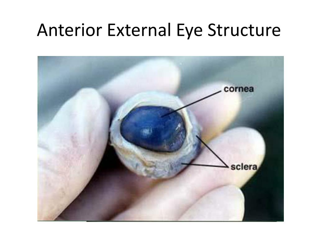
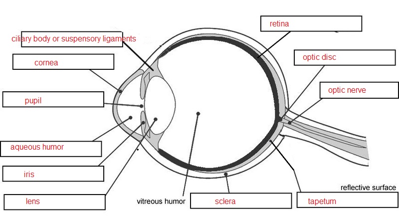
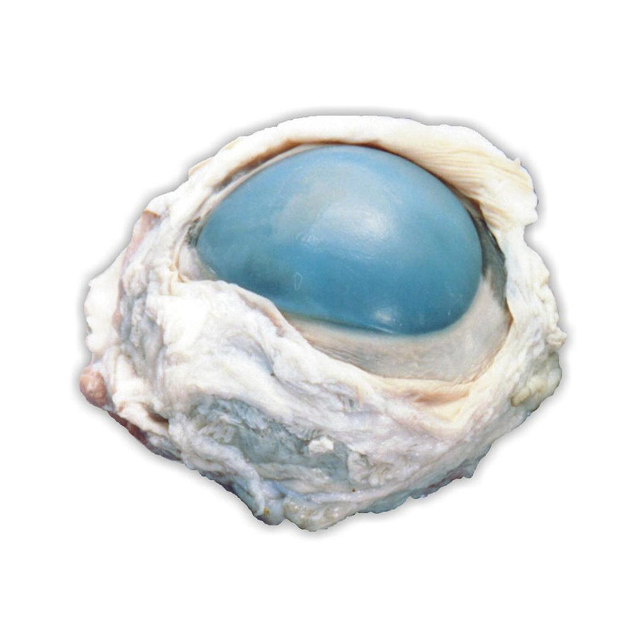
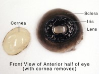
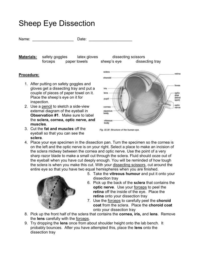
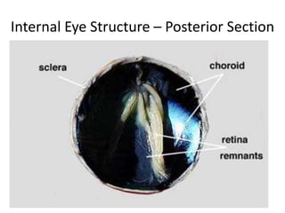
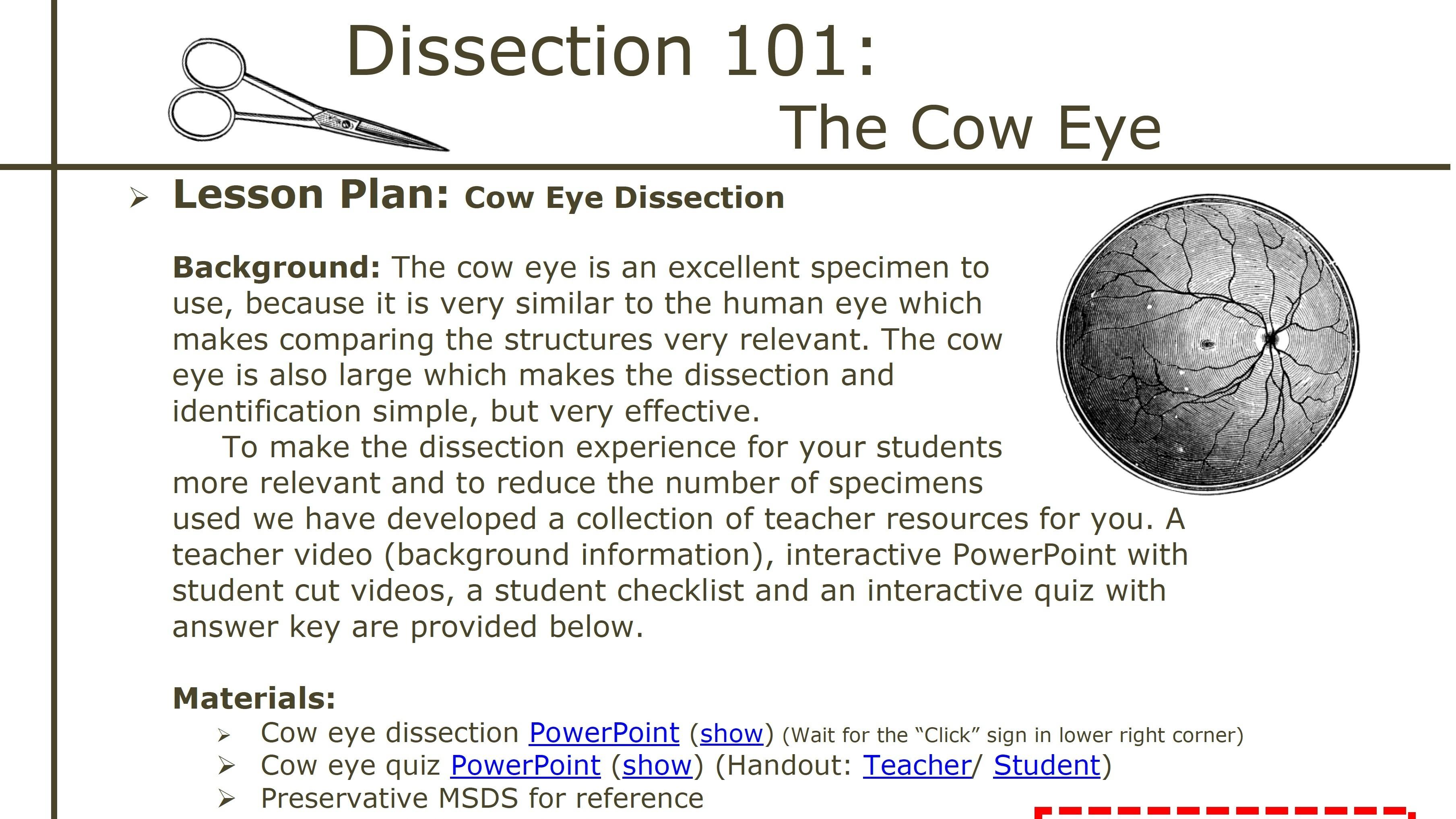
45 cow eyeball dissection labeled
PDF Cow Eye Dissection Lab - Home Science Tools This cow eye dissection kit comes with everything you need to conduct a lab examination. Safety Guidelines • Work in a place separate from eating and food preparation areas. • Use disposable latex gloves or nitrile gloves during the dissection and cleanup. • Use only dissection tools provided. [Solved] How to label a cow eye dissection? | Course Hero One way is to use a permanent marker to label the different parts of the eye. Another way is to use a piece of tape and write the labels on the tape. You can also use labels that are made specifically for eye dissections. When labeling a cow eye dissection, the most essential thing to remember is to be precise.
Cow Eye Dissection - YouTube About Press Copyright Contact us Creators Advertise Developers Terms Privacy Policy & Safety How YouTube works Test new features Press Copyright Contact us Creators ...
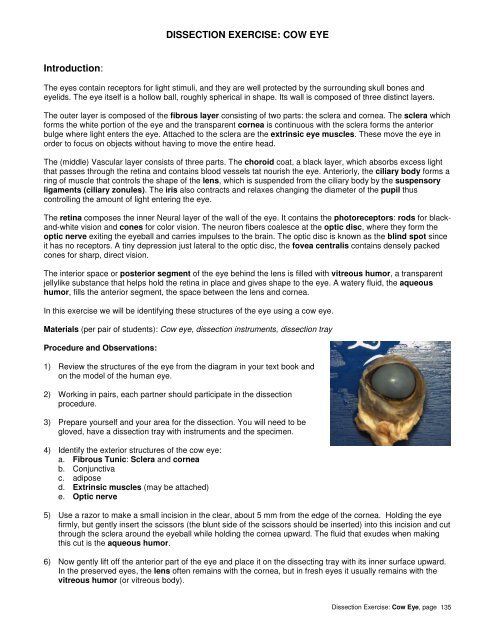
Cow eyeball dissection labeled
Lab: Cow Eye Dissection Flashcards | Quizlet the white outer layer of the eyeball. At the front of the eye it is continuous with the cornea aqueous humor transparent, watery fluid similar to plasma, but containing low protein concentrations. It is secreted from the ciliary epithelium, a structure supporting the lens vitreous humor Cow Eye Dissection Teaching Resources | Teachers Pay Teachers This is a comprehensive dissection guide of the cow eye, designed for a high school or early college Biology or Anatomy & Physiology class. The guide includes step-by-step instructions and labeled diagrams that will lead students through the external anatomy of the eye, followed by dissection of the internal structures. Cow Eye Dissection & Parts of the Eye Diagram | Quizlet cornea Clear, outer layer of the front of the eye. sclera White, outermost layer of the eye. Helps maintain shape and gives attachment to muscles. photoreceptors The cells in the retina that respond to light (rods and cones) rods Photoreceptor cells in the eye that detect black, white, and gray cones Photoreceptor cells in the eye that detect color
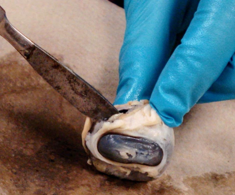
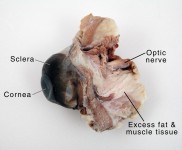
Cow eyeball dissection labeled. Cow Eye Dissection & Anatomy Project | HST Learning Center Cow Eye Dissection: Internal Anatomy 1. Place the cow's eye on a dissecting tray. The eye most likely has a thick covering of fat and muscle tissue. Carefully cut away the fat and the muscle. As you get closer to the actual eyeball, you may notice muscles that are attached directly to the sclera and along the optic nerve. Cow Eyeball Dissection - YouTube Mmm..juicy, juicy eyeballs! Join your guides, "Lefty" and "Righty" once more, as they look deeply into the eyes of a cow that's given its all during this An... Cow Eye for Dissection Specimen - Home Science Tools The economy classroom cow eye dissection pack is great for smaller classrooms or for students to share. This is a plain preserved specimen of a single adult cow eye. 10+ pricing is based on bulk-packed specimens. When necessary, you will receive a combination of 10-packs and individually packed specimens to fill your order. Cow Eye Dissection Guide - Google Slides Cow Eye. Use the point of a scissors or a scalpel to make an incision through the layers of the eye capsule (similar to figure 1); there are three layers from the exterior: sclera, whitish/grey, continuous with the transparent cornea, choroid, thin dark black layer and the retina, thin greyish/pink layer. Use a scissors to dissect the entire ...
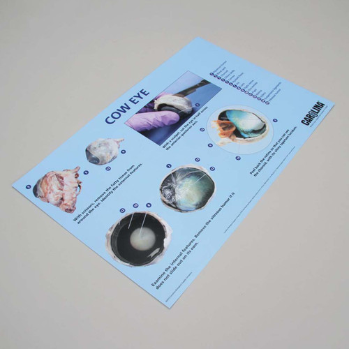
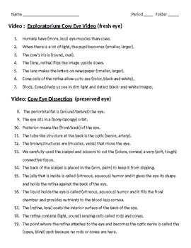
Cow Eye Dissection | Carolina.com Dissection Supplies We offer a full range of dissecting equipment to fit all your lab needs. There are sets available for all skill levels or can be customized. Lab Equipment Carolina is your quality source for a well-equipped lab. Take time to view our high quality science lab equipment that has proven durability to handle any lab activity. Cow Eye Dissection Lab from Anatomy and Physiology The Cow Eye Dissection Lab. The mammalian eye consists of many specialized cells and tissues that make up several different structures. The structures have certain functions and together, they form images that are interpreted by the brain. In this investigation, you identify the structures of a cow eye and learn their functions. Cow Eyeball Dissection Tutorial with Anatomy & Physiology ... - YouTube #baldguysci #edchat #kicksomeclass #dissection #anatomyphysiology #science #sheepeyeIn this cow eye dissection tutorial, we will explore the basic eyeball an... anatomy and physiology of cow anatomy and physiology of cow anatomy and physiology of cow Sheep brain labeled anatomy external lobe dissection frontal physiology nervous system occipital spinal cord savalli. Cow eye dissection labeled. Sheep heart anatomy and physiology of cow
Cow Eye Dissection & Parts of the Eye Diagram | Quizlet cornea Clear, outer layer of the front of the eye. sclera White, outermost layer of the eye. Helps maintain shape and gives attachment to muscles. photoreceptors The cells in the retina that respond to light (rods and cones) rods Photoreceptor cells in the eye that detect black, white, and gray cones Photoreceptor cells in the eye that detect color Cow Eye Dissection Teaching Resources | Teachers Pay Teachers This is a comprehensive dissection guide of the cow eye, designed for a high school or early college Biology or Anatomy & Physiology class. The guide includes step-by-step instructions and labeled diagrams that will lead students through the external anatomy of the eye, followed by dissection of the internal structures. Lab: Cow Eye Dissection Flashcards | Quizlet the white outer layer of the eyeball. At the front of the eye it is continuous with the cornea aqueous humor transparent, watery fluid similar to plasma, but containing low protein concentrations. It is secreted from the ciliary epithelium, a structure supporting the lens vitreous humor
















Post a Comment for "45 cow eyeball dissection labeled"