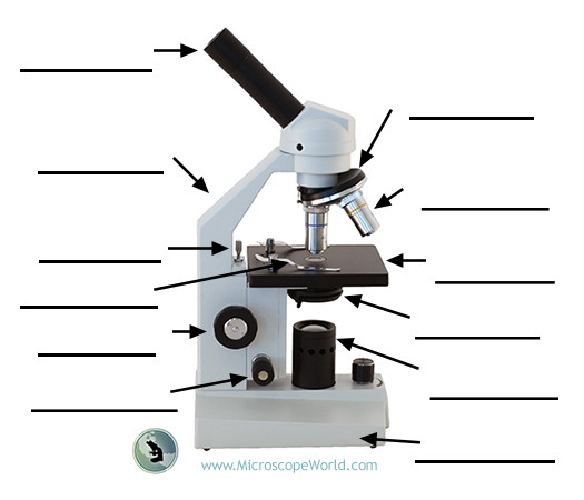
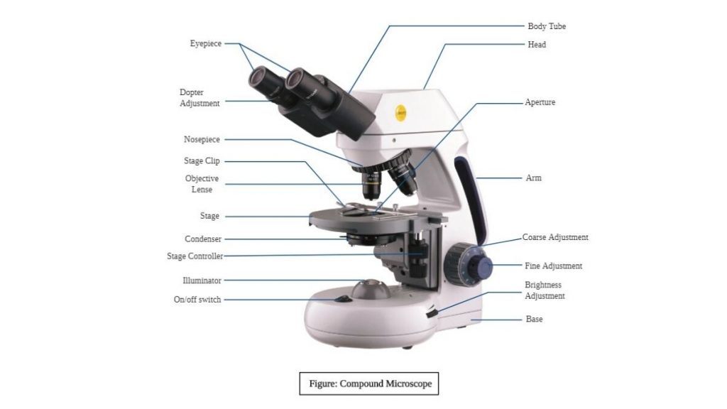
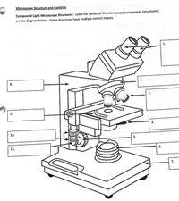
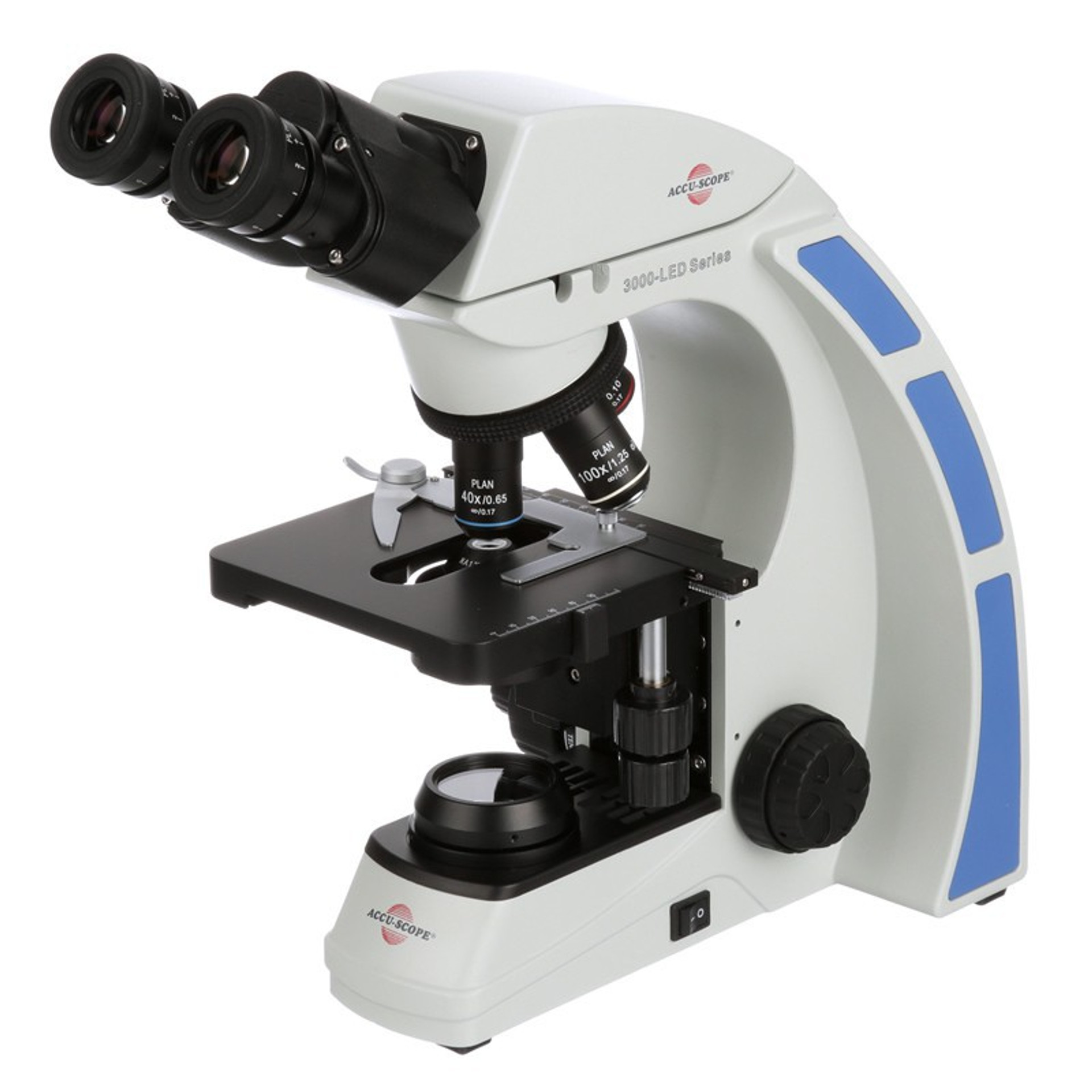
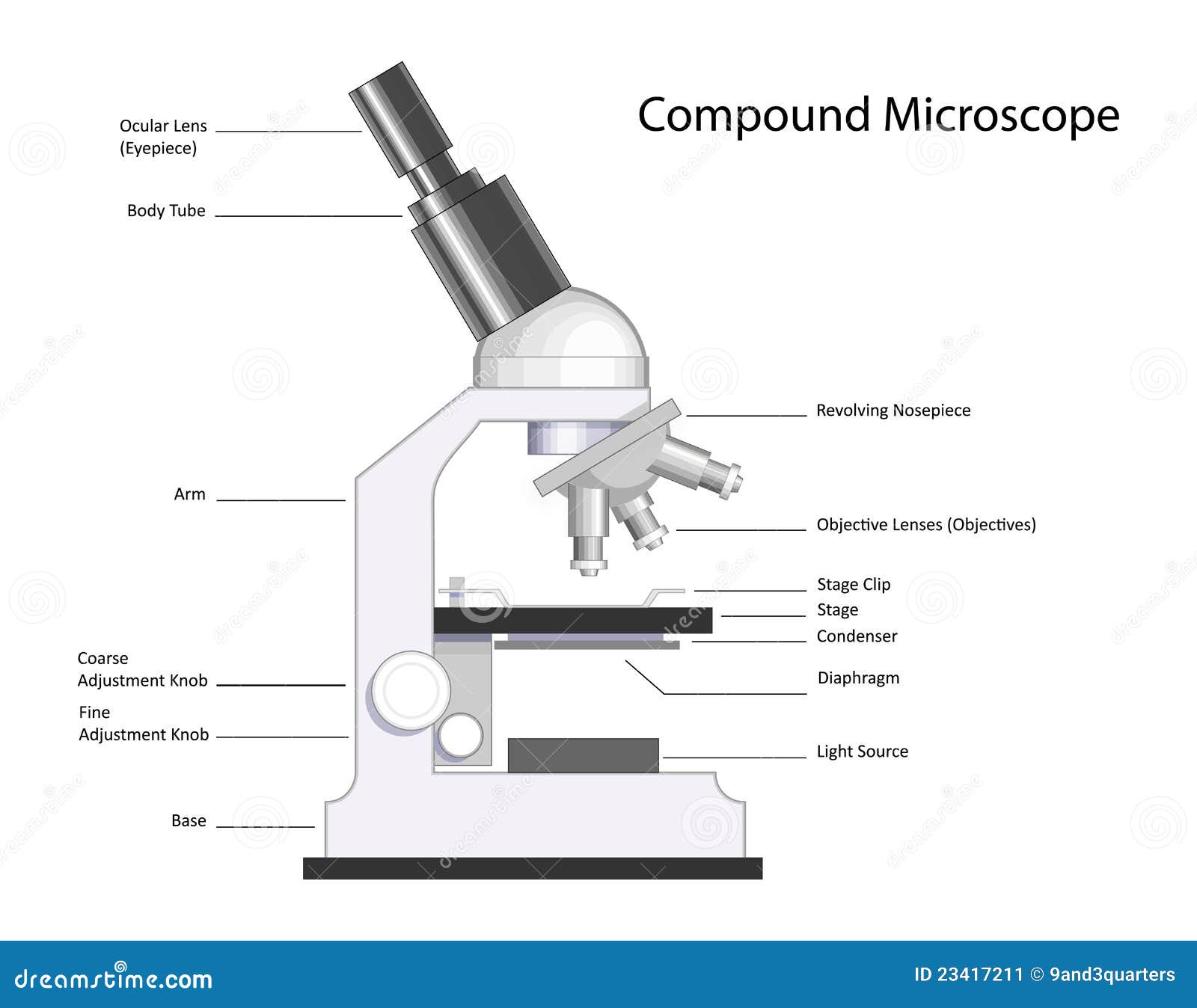
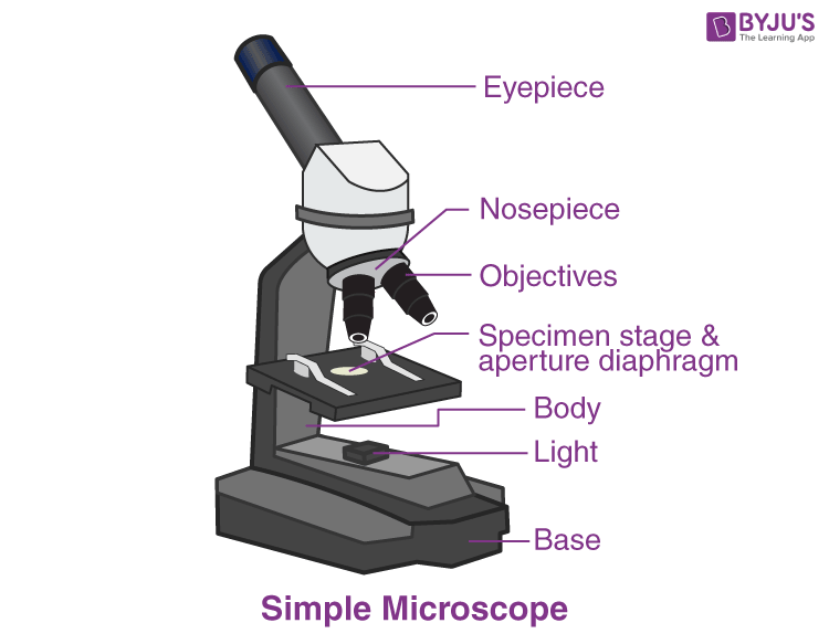
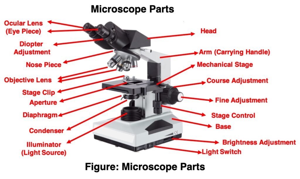
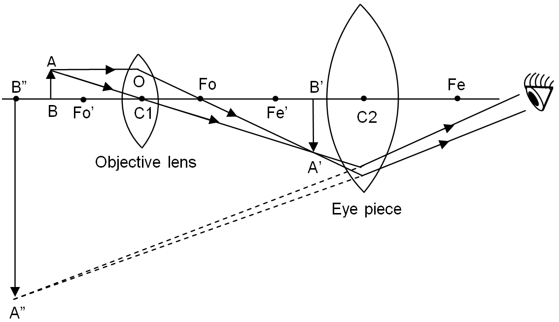
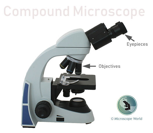
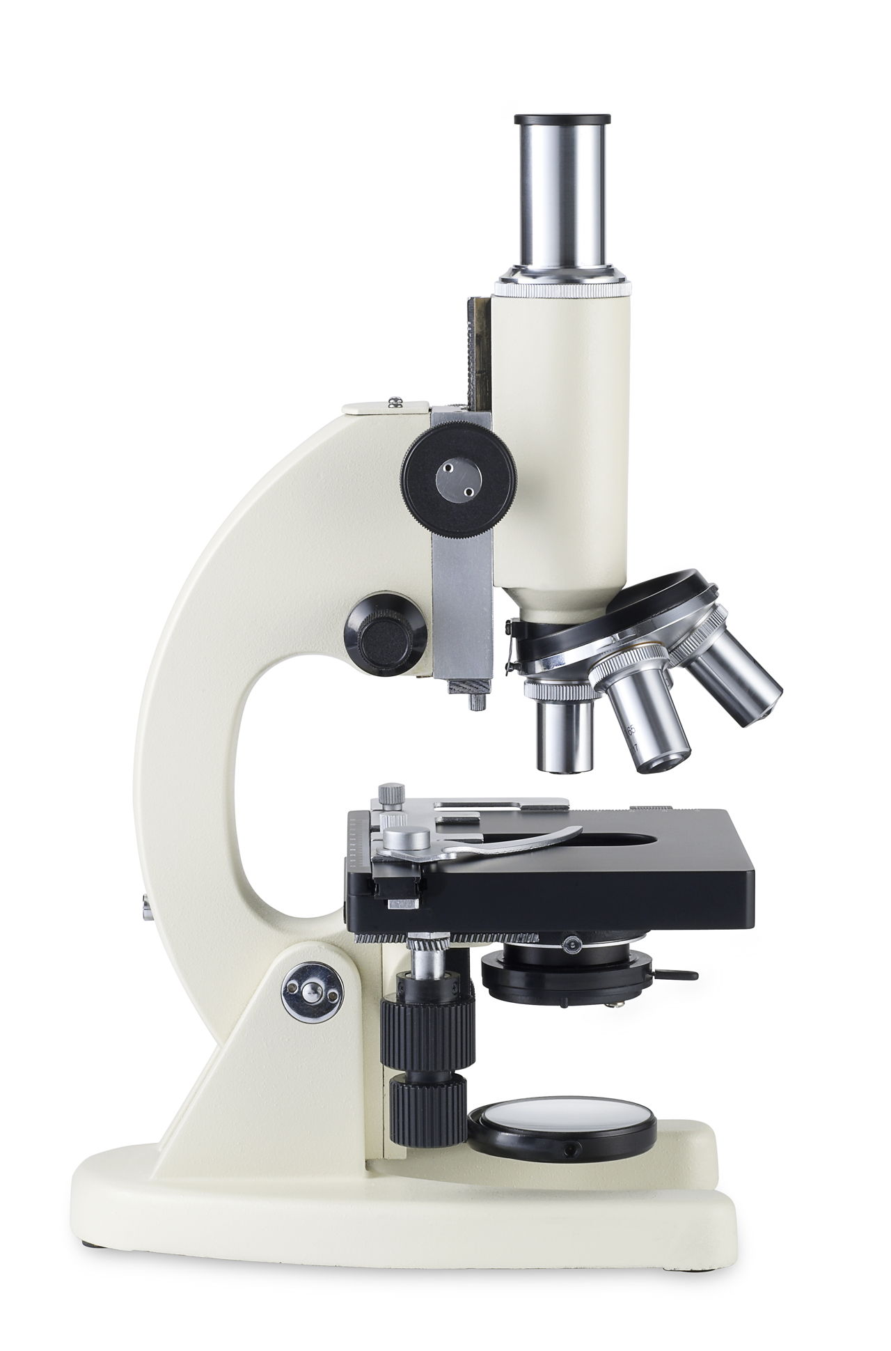
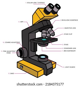
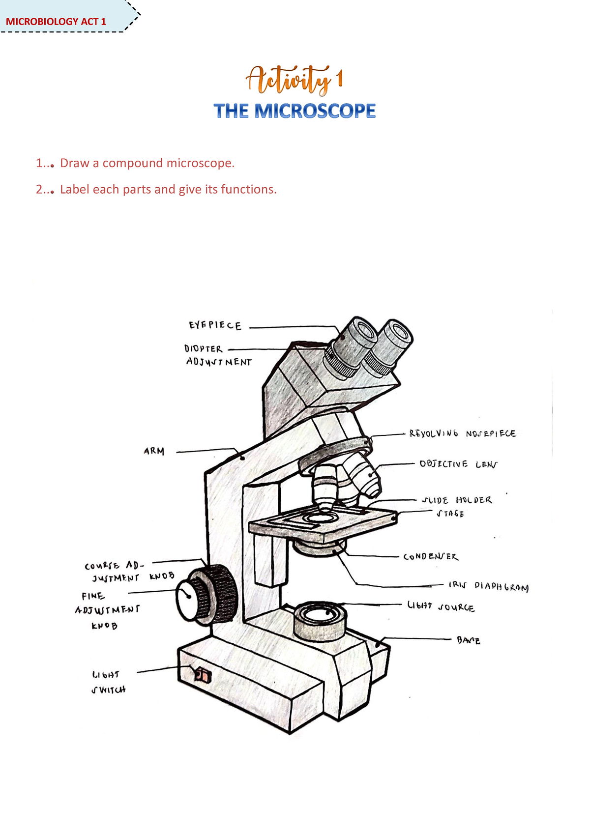
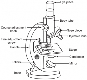
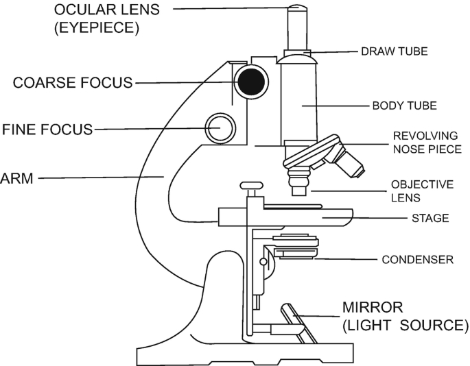
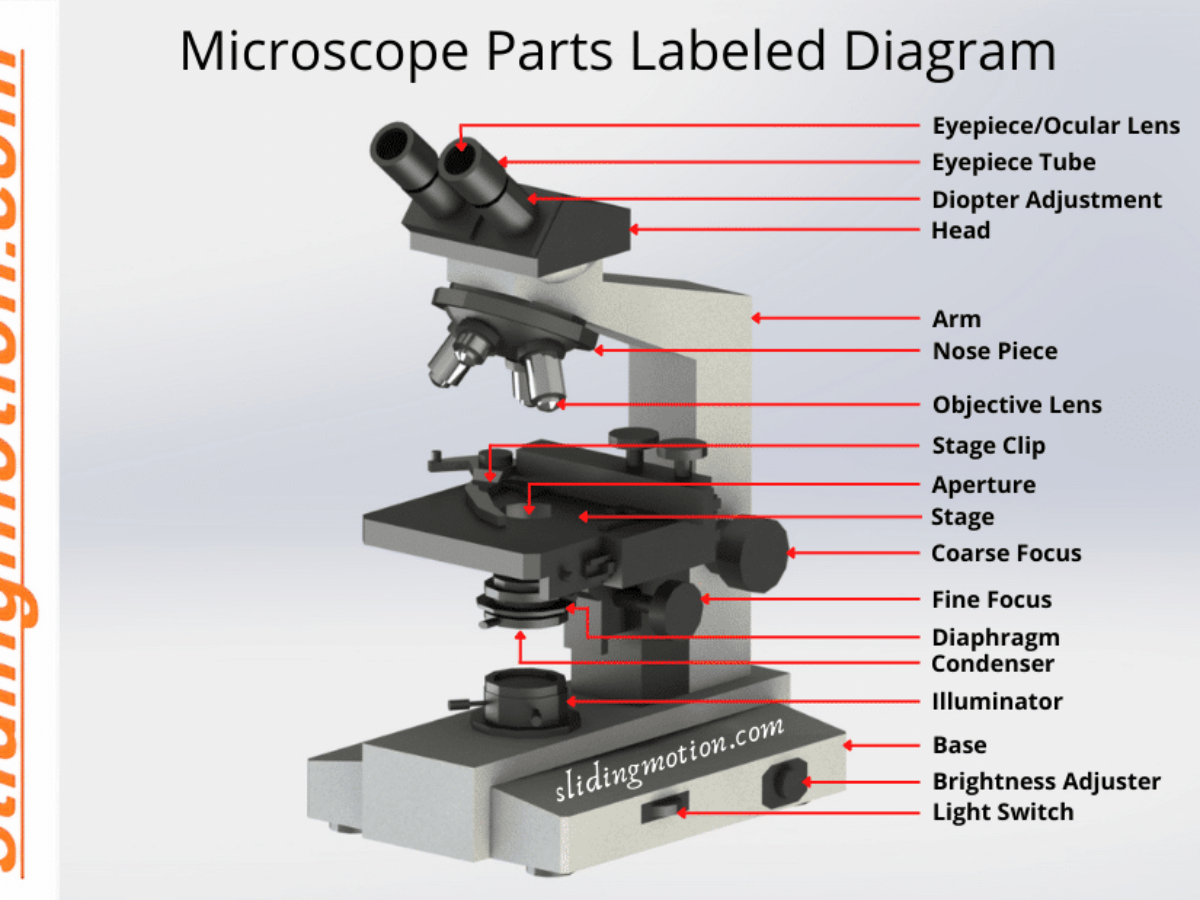
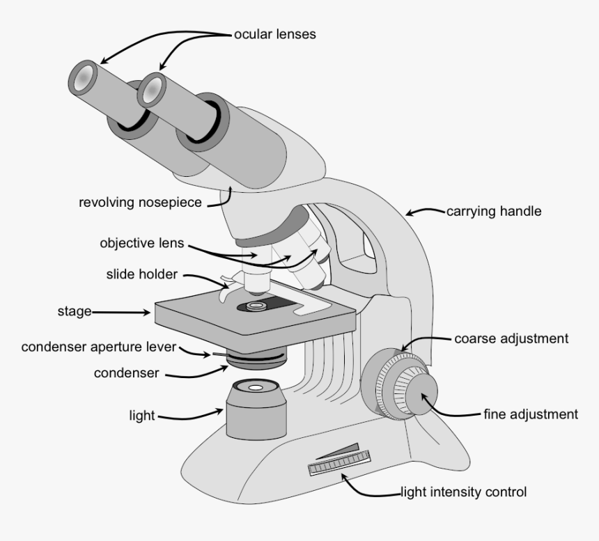
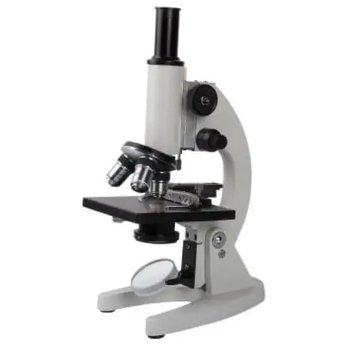
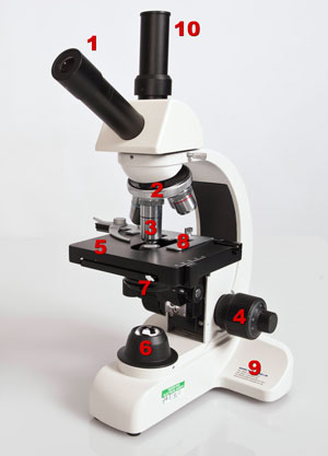
45 compound microscope diagram with labels
rsscience.com › stereo-microscopeParts of Stereo Microscope (Dissecting microscope) – labeled ... If you would like to learn optical components of a compound microscope, please visit Compound Microscope Parts – Labeled Diagram and their Functions, and this article. How to use a stereo (dissecting) microscope. Follow these steps to put your stereo microscopes in work: 1. Set your microscope on a tabletop or other flat sturdy surface where ... en.wikipedia.org › wiki › Glossary_of_geneticsGlossary of genetics - Wikipedia Also three-prime untranslated region and trailer sequence. 3'-end Also three-prime end. One of two ends of a single linear strand of DNA or RNA, specifically the end at which the chain of nucleotides terminates at the third carbon atom in the furanose ring of deoxyribose or ribose (i.e. the terminus at which the 3' carbon is not attached to another nucleotide via a phosphodiester bond ; in ...
› articles › s41578/019/0121-4High-entropy alloys | Nature Reviews Materials Jun 18, 2019 · High-entropy alloys have greatly expanded the compositional space for alloy design. In this Review, the authors discuss model high-entropy alloys with interesting properties, the physical ...
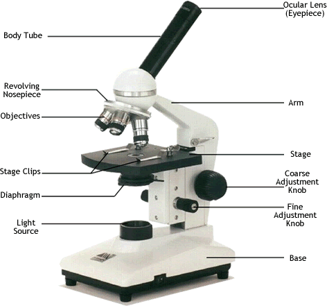
Compound microscope diagram with labels
en.wikipedia.org › wiki › FluorescenceFluorescence - Wikipedia The chemical compound responsible for this fluorescence is matlaline, which is the oxidation product of one of the flavonoids found in this wood. [1] In 1819, Edward D. Clarke [5] and in 1822 René Just Haüy [6] described fluorescence in fluorites , Sir David Brewster described the phenomenon for chlorophyll in 1833 [7] and Sir John Herschel ... › public › staticpagesGuide to Pipetting - Pipettes and Pipette Tips Notice: 1.) To get accurate results, calibrate the pipette with the volatile compound you want to pipette. If you use air displacement pipettes, aspirate and dispense the liquid a few times keeping the tip in the liquid. By doing so, the air inside the pipette will be saturated with vapor of the volatile compound. 2.) › techniques › multi-photonMultiphoton Microscopy | Nikon’s MicroscopyU This is a valuable enhancement to the capability of the conventional microscope since ultraviolet wavelengths below approximately 300 nanometers are very problematic for regular microscope optics. Higher-order non-linear effects, such as four-photon absorption, have been experimentally demonstrated, although it is unlikely that these phenomena ...
Compound microscope diagram with labels. microscopeinternational.com › compound-microscopeCompound Microscope Parts, Functions, and Labeled Diagram Nov 18, 2020 · The individual parts of a compound microscope can vary heavily depending on the configuration & applications that the scope is being used for. Common compound microscope parts include: Compound Microscope Definitions for Labels Eyepiece (ocular lens) with or without Pointer: The part that is looked through at the top of the compound microscope ... › techniques › multi-photonMultiphoton Microscopy | Nikon’s MicroscopyU This is a valuable enhancement to the capability of the conventional microscope since ultraviolet wavelengths below approximately 300 nanometers are very problematic for regular microscope optics. Higher-order non-linear effects, such as four-photon absorption, have been experimentally demonstrated, although it is unlikely that these phenomena ... › public › staticpagesGuide to Pipetting - Pipettes and Pipette Tips Notice: 1.) To get accurate results, calibrate the pipette with the volatile compound you want to pipette. If you use air displacement pipettes, aspirate and dispense the liquid a few times keeping the tip in the liquid. By doing so, the air inside the pipette will be saturated with vapor of the volatile compound. 2.) en.wikipedia.org › wiki › FluorescenceFluorescence - Wikipedia The chemical compound responsible for this fluorescence is matlaline, which is the oxidation product of one of the flavonoids found in this wood. [1] In 1819, Edward D. Clarke [5] and in 1822 René Just Haüy [6] described fluorescence in fluorites , Sir David Brewster described the phenomenon for chlorophyll in 1833 [7] and Sir John Herschel ...















Post a Comment for "45 compound microscope diagram with labels"